


The urinary bladder is one of the key organs of the excretory system in the human body. While the kidneys are responsible for the production of urine, the bladder is responsible for its storage and final expulsion. The bladder is located in the lower part of the abdomen, in the pubic area – thanks to this specific hiding, it can protect itself from injuries by the surrounding pelvic bones. If the bladder is empty, it takes the shape of a funnel widening at the top and narrowing at the bottom, while if it is full it becomes a spherical form. The capacity of the bladder is largely determined by the anatomy, but generally its capacity is between 0,4 and 0,6 liters.
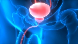
Urinary bladder – anatomy
Ilana ti ito àpòòtọ indicates its innervation and numerous protective layers, protecting against injuries, for example from the pelvic bones. It is built mainly of smooth muscles, connective tissue and blood vessels, in its shape we distinguish the top, shaft, bottom and neck. The walls of the bladder consist of three layers – the first protective layer, the outer, so-called serous membrane, the layer located in the middle – between the outer and inner parts – i.e. the middle layer (muscle tissue) and the inner layer, i.e. the serous membrane. essential element structure of the bladder is its core that it creates detrusor muscle allowing free changes of the shape of the organ in all directions. At the very bottom of the bladder is the urethra, which ultimately expels urine from the human body. For men, the situation is a bit more complicated in this respect, because bladder anatomy assumes that the coil passes through the center of the prostate gland, the so-called prostate. This is the source of many problems in this area in connection with urination. Very often there is an enlargement of the gland and caused by this pressure on the coil. This usually results in a reduced stream intensity, and in more serious cases, inability to urinate completely. A very important element of the structure of the urinary bladder is the urethral sphincter, because it is thanks to it that it is possible to control the excretion of urine. It is a muscle that constantly maintains tension, thanks to which the opening of the urethra is closed during the storage of urine. Its role is particularly useful in situations where there is a sudden increase in pressure in the abdominal area – even during a fit of laughter, coughing, sneezing. Sfincter it can prevent unwanted urine output through natural compression.
Urinary bladder – don’t go without it
The human body works in such a way that it naturally accumulates urine and then excretes it. It is the organ that helps in fulfilling these functions àpòòtọ. It allows you to store the filtered liquid, and thanks sphincter keeping it under control. Ultimately, it’s work àpòòtọ causes urine to be expelled. The centers supervising these activities are located in the nervous system – in the cerebral cortex, spinal cord, in the peripheral ganglia. This is where the signals come in bladder filling up. Capacity àpòòtọ for it is not unlimited. If the fluid fills it in 1/3, then signals flow from the receptors of the bladder walls directly to the cerebral cortex, which signals the need to defecate. If the person then does not react and does not urinate, these signals gain in strength, resulting in a feeling of intense, sometimes even painful urge. At the same time, work is activated at this very moment urethral sphincterswhich prevent the unwanted excretion of urine. If defecation is finally possible, the nerve centers stop sending alarming blocking signals, sphincter limp and urine is excreted. After bowel movements are complete, the organs contract again, preparing for the next collection of urine in the bladder.