


Awọn akoonu
Aneurysm Ruptured - Itumọ, Awọn ami aisan ati Awọn itọju
The aneurysm is a swelling of the wall of the artery, the rupture of which leads to hemorrhage, with a risk of death. It can involve different organs such as the kidneys, heart or brain.
Definition of aneurysm
An aneurysm is characterized by a hernia in the wall of an artery, resulting in the weakening of the latter. Aneurysms can remain silent or rupture, causing serious health problems or even death.
An aneurysm can occur in large arteries such as those that supply blood to the brain and aorta.
An aneurysm can also occur in peripheral arteries – usually behind the knee – although rupture of these is relatively rare.
The two most important places for aneurysms are:
In the artery that leaves the heart directly: it is an aortic aneurysm. It includes the aneurysm of thethoracic aorta and the aneurysm of theabdominal aorta.
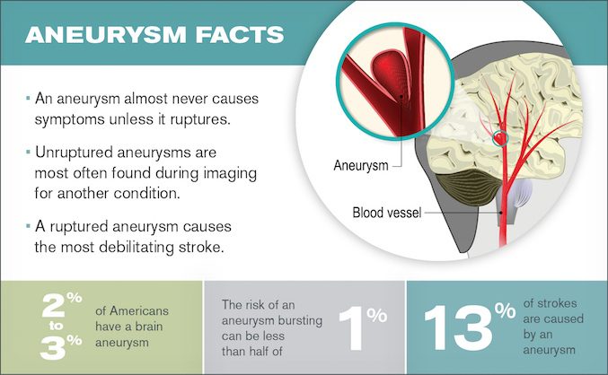
In the artery that supplies the brain: it is a cerebral aneurysm, often called an intracranial aneurysm.
There are other types of aneurysms such as those affecting the mesenteric artery (affecting the artery that feeds the gut) and those affecting the splenic artery and occurring in the spleen.
Concerning the cerebral aneurysm, the latter can cause a leak or a rupture of blood, causing bleeding in the brain: one speaks then ofọpọlọ hemorrhagic type. Most often a brain aneurysm from a ruptured vessel occurs in the space between the brain and the tissues (meninges) covering the brain. This type of hemorrhagic stroke is called a subarachnoid hemorrhage. Most brain aneurysms, however, do not rupture. Brain aneurysms are more common in adults than in children and more common in women than in men.
Causes of ruptured aneurysm
How are aneurysms formed?
Swelling in an artery occurs as a result of thinning of its wall, which allows blood pressure to abnormally widen the arterial wall.
An aortic aneurysm usually takes the form of a bulge that is uniform all around the artery, whereas the cerebral aneurysm instead results in the formation of a bulge that takes the shape of a sac, usually at a place where the arteries are most fragile.
Ruptured brain aneurysms are the most common cause of a type of stroke known as subarachnoid hemorrhage. This type of stroke is less common than ischemic stroke.
Why do aneurysms develop?
It is not completely understood why the arterial wall weakens and how it does to cause an aneurysm.
It is known, however, that there are a number of risk factors (see below) that are known to be associated with the development of aneurysms.
Diagnosis of brain aneurysm
If you have a sudden or severe headache or other symptoms possibly related to an aneurysm, you will have a test or series of tests to determine if you are bleeding into the space between your brain and surrounding tissue (hemorrhage subarachnoid) or a form of stroke.
If bleeding has occurred, the emergency team will determine if an aneurysm is the cause.
If you have symptoms of a non-rupturing brain aneurysm – such as pain behind your eye, vision problems, and paralysis on one side of your face – you will likely undergo the same tests.
Diagnostic tests include:
- Computerized tomography (CT). This CT scan is usually the first test used to determine if there is bleeding in the brain.
- Magnetic resonance imaging (MRI). An MRI uses a magnetic field and radio waves to create detailed images of the brain. She assesses the arteries in detail can identify the site of the aneurysm.
- Cerebrospinal fluid test. Subarachnoid hemorrhage often leads to the presence of red blood cells in the cerebrospinal fluid (fluid surrounding the brain and spine). This test is done if there are symptoms of aneurysm.
- Cerebral angiography or angioscanner. During this procedure, the doctor injects a dye into a catheter in a large artery – usually in the groin. This test is more invasive than others and typically used when other diagnostic tests do not provide enough information.
Using imaging tests to screen for unruptured brain aneurysms is generally not recommended unless the patient has a family history with a first-degree relative (parent, sibling).
Complications of the aneurysm
The majority of people living with an aneurysm do not suffer from the complications. Managing risk factors is important, however.
Complications of the aneurysm are as follows:
- Venous thromboembolism: Blockage of a vein by a blood clot can cause pain in an organ such as the abdomen or brain, and in the latter case can cause a stroke.
- Severe chest and / or lumbar pain: it occurs following a silent or ruptured aortic aneurysm.
- Ikọju Angina : Certain types of aneurysm can lead to angina pectoris, pain related to narrowed arteries that provide poor supply to the heart.
The case of cerebral aneurysm
When a brain aneurysm ruptures, the bleeding usually only lasts a few seconds. The bleeding can cause damage to surrounding brain cells (neurons). It also increases the pressure inside the skull.
If the pressure gets too high, the blood and oxygen supply to the brain can be disrupted to the point that unconsciousness or even death can occur.
Complications that can develop after an aneurysm ruptures include:
- Another bleeding. A ruptured aneurysm may bleed again, causing further damage to brain cells.
- Vasospasm. Following an aneurysm, the blood vessels in the brain can narrow suddenly and temporarily: this is vasospasm. This abnormality can restrict blood flow to brain cells, causing ischemic stroke and causing further damage to neurons.
- Hydrocephalus. When a ruptured aneurysm causes bleeding into the space between the brain and surrounding tissue (subarachnoid hemorrhage), the blood can block the flow of fluid (called cerebrospinal fluid) surrounding the brain and the body. opa eyin. This condition can cause an excess of cerebrospinal fluid which increases the pressure on the brain and can damage tissues: it is hydrocephalus.
- Hyponatremia. Subarachnoid hemorrhage following a cerebral aneurysm can disrupt the sodium balance in the blood. This can produce damage in the hypothalamus, an area at the base of the brain. A awọn ipele iṣuu soda kekere ninu ẹjẹ (called hyponatremia) can lead to swelling of neurons and permanent damage.